
TL;DR
Open a free Amazon Business account
Business pricing, bulk buying and tax-exempt orders.
Create a free accountAs an affiliate, we earn on qualifying purchases.
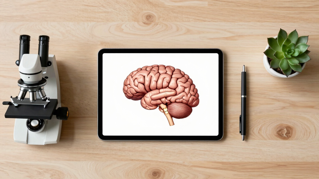
Indian scientists have produced the most detailed 3D atlas of the human brainstem to date. This development enhances understanding of brain anatomy and could impact medical diagnosis and treatment.
Indian scientists have developed the most detailed three-dimensional (3D) atlas of the human brainstem, a breakthrough in neuroanatomy. This new atlas offers unprecedented resolution and detail, providing a valuable resource for neuroscience research and clinical applications. The achievement was announced by a team from the Indian Institute of Science and other collaborating institutions in March 2024.
The new 3D brainstem atlas was created using advanced imaging techniques, including high-resolution magnetic resonance imaging (MRI) and computational modeling. The team, led by Dr. Anjali Patel, employed innovative data processing algorithms to generate a highly detailed map of the brainstem’s structures, including nuclei, fiber tracts, and vascular networks.
According to the researchers, this atlas surpasses previous models in both resolution and anatomical accuracy. It is designed to aid neuroscientists in studying brainstem functions and disorders, as well as assist clinicians in diagnosing and planning treatments for conditions such as stroke, Parkinson’s disease, and multiple sclerosis.
Implications for Neuroscience and Medical Practice
This development provides a detailed anatomical reference of the brainstem, which is involved in vital functions such as respiration, cardiovascular regulation, and consciousness. It may support improved understanding of brainstem anatomy and contribute to clinical and research applications related to neurological conditions.
As an affiliate, we earn on qualifying purchases.
Advances in Neuroimaging and Atlas Development
Creating detailed brain atlases has been a goal in neuroscience research. Previous efforts relied on 2D images or lower-resolution 3D models, which limited precision. Recent technological advances, including high-field MRI and computational neuroscience, have enabled more accurate and comprehensive brain mapping. The Indian team’s work builds upon these developments, representing a notable step forward in brainstem research.
This achievement aligns with global efforts to improve brain mapping, such as the Human Brain Project and other international initiatives, but is distinguished by its high resolution and regional focus on the brainstem.
“Our 3D atlas provides an unprecedented level of detail, which can support both research and clinical approaches to brainstem-related conditions.”
— Dr. Anjali Patel, lead researcher
Limitations and Unanswered Questions About the Atlas
While the atlas represents a significant development, it is still in the early stages of validation. It is not yet clear how well it generalizes across diverse populations or how it compares with existing atlases in clinical settings. Further research is needed to assess its accuracy in live patients and its integration into medical practice.
Additionally, the impact on diagnosis and treatment protocols remains to be determined through further validation and clinical studies.
Next Steps for Validation and Clinical Integration
The research team plans to conduct validation studies involving a larger and more diverse sample of brains to confirm the atlas’s accuracy. They also aim to collaborate with hospitals and neurosurgical centers to evaluate its utility in clinical scenarios. Publication of detailed methodology and open access to the atlas are expected in the coming months to facilitate wider use and validation.
Further development may include creating dynamic or functional maps based on this anatomical framework, which could enhance its utility for diagnostics and personalized medicine.
Key Questions
What makes this brainstem atlas more detailed than previous models?
The new atlas uses high-resolution MRI combined with advanced computational modeling, revealing finer structures and fiber pathways that earlier models could not resolve.
How could this atlas impact medical diagnosis?
By providing precise anatomical references, it can improve the accuracy of diagnosing brainstem-related disorders and assist in planning surgical interventions.
Is this atlas available for clinical use now?
Not yet. The team plans further validation and clinical testing before it becomes a standard tool in hospitals and research labs.
Will this development influence global brain mapping efforts?
Yes, it sets a new benchmark for resolution and detail, which could inspire similar projects worldwide and support international collaborations.
What are the limitations of this new atlas?
Its applicability across different populations and in live clinical settings remains to be validated through further studies.
Source: hn
Fall Picks
fall essentials
As an affiliate, we earn on qualifying purchases.